Home
/ Compact Bone Diagram Microscope : Compact Bone Tissue Images Stock Photos Vectors Shutterstock : From the spongy bone histology slide, we will identify the following important histological features under the light compound microscope.
Compact Bone Diagram Microscope : Compact Bone Tissue Images Stock Photos Vectors Shutterstock : From the spongy bone histology slide, we will identify the following important histological features under the light compound microscope.
Compact Bone Diagram Microscope : Compact Bone Tissue Images Stock Photos Vectors Shutterstock : From the spongy bone histology slide, we will identify the following important histological features under the light compound microscope.. Learn vocabulary, terms, and more with flashcards, games, and other study tools. The marrow in these images is red marrow. This human bone section shows the haversian canal (or osteon) structure of compact bone tissue. 2 compact bone we know that compact bone is very dense it is also very complex when viewed under a microscope. Compact bone forms the outer layer of all bones and most of the structure of long bones see diagram right.
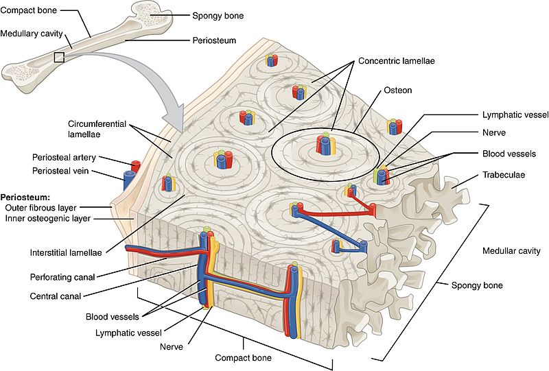
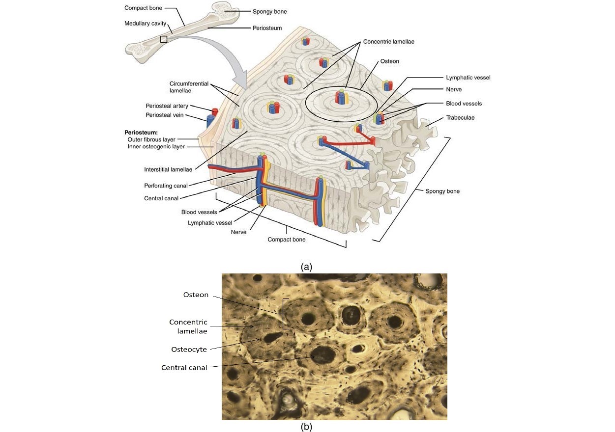
The osteon consists of a central canal called the osteonic (haversian) canal, which is surrounded by concentric rings (lamellae) of. Spongy bone diagram schematic diagram. (b) in this micrograph of the osteon, you can clearly see the concentric lamellae and central canals. The cells of compact bone, which is also called cortical bone, appear to be tightly packed into a solid mass. This human bone section shows the haversian canal (or osteon) structure of compact bone tissue.
14 4 Structure Of Bone Biology Libretexts from bio.libretexts.org A diagram of the anatomy of a bone, showing the compact bone. Each osteon looks like a ring with a light spot in the center. Compact bone diagram osteon compact bone ap pinterest anatomy human anatomy and. Start studying microscopic anatomy of compact bone part 2. The remainder is cancellous bone, which has a spongelike appearance with numerous large spaces and is found in the. The trabeculae are only a few cell layers thick. The cells of compact bone, which is also called cortical bone, appear to be tightly packed into a solid mass. Best worksheet compact bone microscope slide labeled free download file 624 diagram of compact bone new jpg wikimedia commons.
In this article we will discuss about the structure of nucleus with the help of suitable diagrams.
Identify structures and functions of the microscopic structure of compact and spongy bone 3. The remainder is cancellous bone, which has a spongelike appearance with numerous large spaces and is found in the. From the spongy bone histology slide, we will identify the following important histological features under the light compound microscope. Compact bone is formed in concentric circles. Spongy bone diagram schematic diagram. Under the microscope, bone can be divided into two types compact bone forms the outer 'shell' of bone. Human bone cross section microscope.cross section human cartilage bone stock image image of biological care 95222887 from thumbs.dreamstime.com. The spaces between the trabeculae contain red or yellow marrow, depending on a person's age and on which bone it is. Diagramme schnell und einfach erstellen. Explain the role of bone salts and the organic matrix in making bone both hard and flexible. Compact bone cross section courtesy: Compact bone diagram osteon compact bone ap pinterest anatomy human anatomy and. The trabeculae are only a few cell layers thick.
Under the microscope, bone can be divided into two types compact bone forms the outer 'shell' of bone. In this article we will discuss about the structure of nucleus with the help of suitable diagrams. From the spongy bone histology slide, we will identify the following important histological features under the light compound microscope. You can think of compact bone as being very similar. Each osteon looks like a ring with a light spot in the center.
5 3 Bone Structure Medicine Libretexts from med.libretexts.org Microscopic structure of bone diagram. Of bone figure 5.3 page 116 objectives 1. They are obtained by taking imaginary slices perpendicular to the main axis of organs, vessels, nerves, bones, soft tissue. 2 compact bone we know that compact bone is very dense it is also very complex when viewed under a microscope. The trabeculae are only a few cell layers thick. This human bone section shows the haversian canal (or osteon) structure of compact bone tissue. Learn vocabulary, terms, and more with flashcards, games, and other study tools. Under the microscope, bone can be divided into two types compact bone forms the outer 'shell' of bone.
There are small canals that run through the bone, which allow blood vessels to penetrate it.
The light spot is a canal that carries a blood vessel and a nerve fiber. Learn vocabulary, terms, and more with flashcards, games, and other study tools. Human bone cross section microscope.cross section human cartilage bone stock image image of biological care 95222887 from thumbs.dreamstime.com. Compact bone cross section courtesy: Spongy bone diagram schematic diagram. In this article we will discuss about the structure of nucleus with the help of suitable diagrams. 2 compact bone we know that compact bone is very dense it is also very complex when viewed under a microscope. Each osteon looks like a ring with a light spot in the center. Compact bone histology slide structure with diagram. Do you want to learn the details of the histology of compact bone with labelled diagram and authentic slide images? The compact bone is composed of calcified extracellular material the bone matrix and 3 major cell types which are osteoblast which ssynthesize and secrete the organic components of bone matrix which include type 1 collagen fibers proteoglycans and several glycoproteins such as ostepnectin. Each bone is a complex living organ that is made up of many cells protein fibers and minerals. (b) in this micrograph of the osteon, you can clearly see the concentric lamellae and central canals.
Under magnification you can clearly see the system of concentric circles that forms compact bone. 111 is generally a round body occupying the centre of the cell. The outlined area is a cross section of an osteon of compact bone. This video describes the microscopic anatomy of compact bone. (b) in this micrograph of the osteon, you can clearly see the concentric lamellae and central canals.
Microscopic Anatomy Of A Tissue Foundations Of Periodontics Microscopic Anatomy Of Compact Bone Tissue from www.dent-wiki.com This human bone section shows the haversian canal (or osteon) structure of compact bone tissue. A photo taken through a microscope that shows the anatomy of compact bone with a detailed view of an osteon. Like other tissues in the body, bones are made up of specialized cells that serve different functions. 0 0000 a shoutout is a way of letting people know of a. The spaces between the trabeculae contain red or yellow marrow, depending on a person's age and on which bone it is. Best worksheet compact bone microscope slide labeled free download file 624 diagram of compact bone new jpg wikimedia commons. If you look at compact bone under the microscope, you will observe a highly organized arrangement of concentric circles that look like tree trunks. The osteon consists of a central canal called the osteonic (haversian) canal, which is surrounded by concentric rings (lamellae) of.
The spaces between the trabeculae contain red or yellow marrow, depending on a person's age and on which bone it is.
Bone tissue is one of the main components of the skeletal system (other components include bone marrow/marrow cavity, collagen fibers etc). 3 mature bone cells, osteocytes, are found in tiny cavities within the matrix called lacunae. (b) in this micrograph of the osteon, you can clearly see the concentric lamellae and central canals. If you look at compact bone under the microscope, you will observe a highly organized arrangement of concentric circles that look like tree trunks. Compact bone histology slide structure with diagram. Learn vocabulary, terms, and more with flashcards, games, and other study tools. 100x on this image you can see several of the structural units of bone tissue (osteons or haversian systems). From the spongy bone histology slide, we will identify the following important histological features under the light compound microscope. The darker ring consists of layers of bone matrix made by cells called. Best worksheet compact bone microscope slide labeled free download file 624 diagram of compact bone new jpg wikimedia commons. The osteon consists of a central canal called the osteonic (haversian) canal, which is surrounded by concentric rings (lamellae) of. 111 is generally a round body occupying the centre of the cell. The marrow in these images is red marrow.
